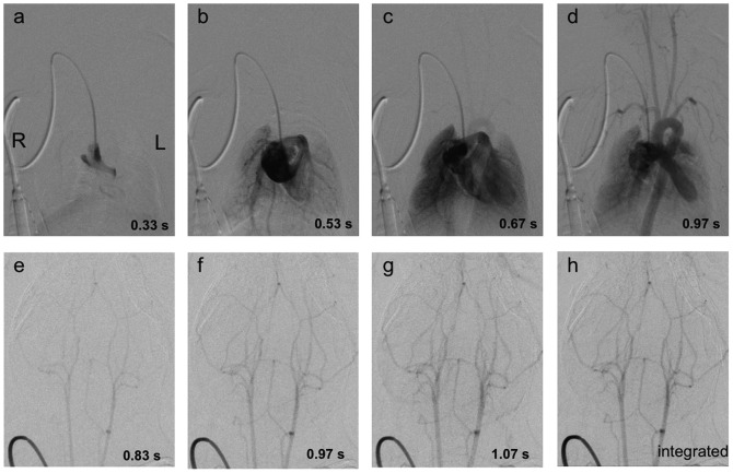
Figure 5. Digital subtraction angiography of a C57/BL6 mouse was performed during injection of 100 µl Iomeprol 300 using the VAMP.
Sequential enhancement of the right atrium (a), the right ventricle and lung arteries (b), the lung parenchyma (c), the left ventricle, the aorta and supra-aortic arteries (d) is shown. A digital subtraction angiography of the cerebral vessels is shown the second row. Images (e)–(g) show the sequential enhancement of the cerebral arteries within one second. Integration of images was performed to improve image quality (h).

