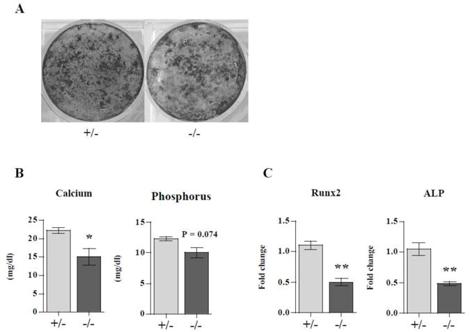
Figure 6. LIMK1−/− osteoblasts have impaired mineralization capacity.

Neonatal calvarial osteoblasts isolated from LIMK1+/− or LIMK1−/− mice were grown to confluence and then treated with 10mM ß-glycerophosphate for 2 weeks. A: Von Kossa staining of cultures from LIMK1+/− and LIMK1−/− mice after 2 weeks of treatment with ß-glycerophosphate. Note reduced staining in the LIMK1−/− cultures. B: Cultures treated with ß-glycerophosphate for 2 weeks after reaching confluence were extracted and calcium phosphorus content quantified or used to prepare RNA for qPCR analyses. Note that the calcium and phosphorus content of the cell layer extracted from the LIMK1−/− mice was lower than in control animals. C: Results of qPCR for runx2 and alkaline phosphatase expression in cell layers of osteoblasts from LIMK1−/− and +/− mice treated for 2 weeks with ß-glycerophosphate. Note that the level of runx2 and alkaline phosphatase expression are significantly lower in LIMK1−/− cultures than in LIMK1+/− cultures. * = p < 0.05; ** = p < 0.01.
