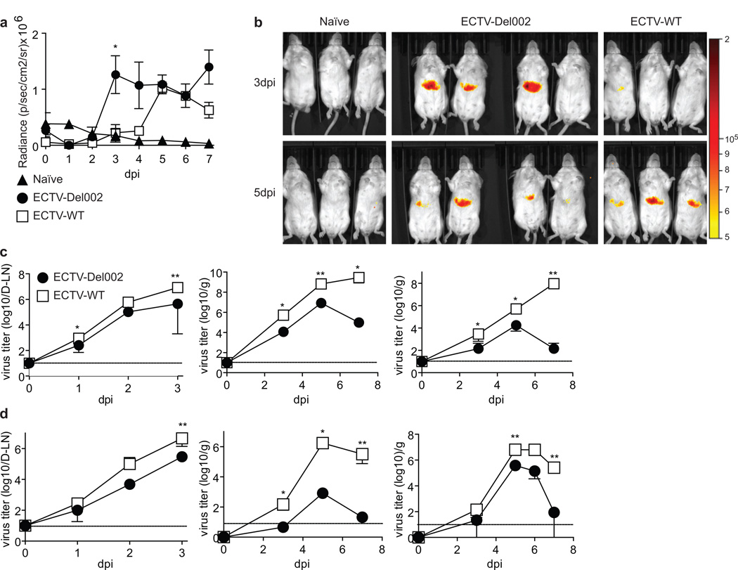
Figure 2. Role of p105bp in NF-kB activation and virus spread.
(a,b) In vivo NF-κB activation: The livers of BALB/c mice were transfected in vivo with an NF-κB reporter plasmid expressing firefly luciferase. 12 days post-transfection the mice were infected with 3,000 pfu ECTV-WT in the footpad and imaged daily for seven days 5 min after luciferin injection. (a) Radiance (p/sec/cm2/sr) in the liver area at the indicated dpi was determined in naïve (n=3), ECTV-Del002-(n=4) and ECTV-WT-infected mice (n=3) and plotted as mean ± SEM with significant difference indicated at 3 dpi. (b) Radiance images of the mice in d at 3 and 5 dpi. (c.d) Virus titers: BALB/c (c) or B6 mice (d) were infected in the footpad with 3,000 pfu of ECTV-WT (empty squares) or ECTV-Del002 (filled circles) viruses. Virus loads were monitored in D-LN (left panels), spleen (middle panels) and liver (right panels). N=5 mice per time point. Data are displayed as mean ± SEM. Horizontal lines indicate limit of detection. See also Figure S2.

