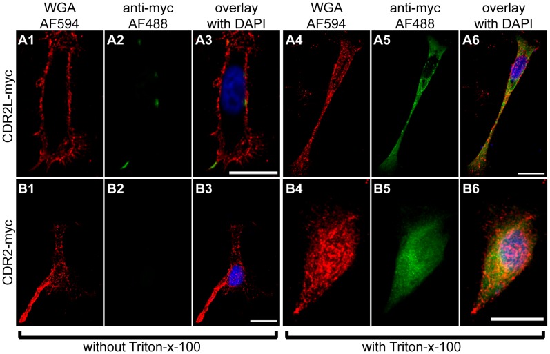
Figure 2. Localization of CDR2L and CDR2 in HeLa cells.
HeLa cells transfected with CDR2L-myc (A1–6) or CDR2-myc (B1–6) were immunocytochemically stained with anti-myc antibodies (green, A2, A5, B2, B5). The membrane was visualized with wheat germ agglutinin (WGA, red, A1, A4, B1, B4). This was performed as a surface staining without addition of Triton-x-100 (A1–3, B1–3) or with Triton-x-100 to permeabilise the membrane (A4–6, B4–6). To show the membrane localisation of CDR2L, only selected slices of a z-stack were projected on top of each other, namely 16 (A1–3) or 23 (A4–6) slices. To show that CDR2 is not localised to the membrane, all slices covering the cells were superimposed, namely 55 (B1–3) and 40 (B4–6) slices. The overlays show that CDR2L can be detected in the membrane (A3), but not CDR2 (B3). Staining of non-permeabilised cells in B2 shows that CDR2 is not membrane-bound. However, the cells were successfully transfected with CDR2 as shown when the cells were permeabilised (B5). The results show that in the permeabilised cells, CDR2L (A5) is localized to the cytoplasm and cellular membrane, whereas CDR2 (B5) is only localised to the cytoplasm and nucleus. Scale bars 20 µm.

