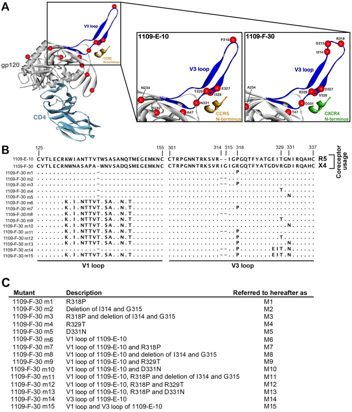
Figure 3. V3 loop alterations segregating X4 from R5 C-HIV Envs, and Env mutagenesis strategy.
(A, left) Ribbon diagram showing a gp120 model of 1109-E-10 Env (grey) in complex with CD4 (light blue) and a sulfated CCR5 N-terminus peptide (orange). The V3 loop is highlighted in blue. The location of amino acids segregating X4 and R5 Envs from this subject shown by space-filled models of their α-carbon atoms (red spheres). (A, center) Close up view of the V3 loop of the R5 1109-E-10 gp120 bound to the CCR5 N-terminus peptide (orange), showing the R5-associated amino acids as red spheres. (A, right) Close up view of the V3 loop of the X4 1109-F-30 gp120 bound to a model of the CXCR4 N-terminus peptide (green), showing the X4-associated amino acids as red spheres. (B) Amino acid sequences of the Env mutants, aligned against the gp120 sequence of the X4 1109-F-30 sequence. Dots indicate residues identical to 1109-F-30, dashes indicate gaps. Numbers refer to amino acid positions in the V1 and V3 loop regions. (C) Brief descriptions of the Env mutants, which are provided in greater detail in Materials and Methods.

