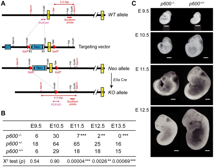
Figure 1. Disruption of p600 results in growth retardation and lethality during embryonic development.
(A) Targeting strategy of p600 locus. The exon encoding the initiating methionine codon of p600 allele was replaced with a Neo cassette by homologous recombination in ES cell lines. The ES cell lines were microinjected into mouse blastocysts to generate chimeric mice. These chimeras were bred to obtain offspring that are heterozygous for ‘Neo allele’. p600 Neo/+ mice were then crossed with EIIa-Cre transgenic mouse [19] to delete the loxP flanked region, yielding ‘KO allele’. The locations of PCR amplicons used for genotyping are as indicated. Restriction enzyme digestion of genomic DNA with BamHI and SpeI produces the 2.5 and 4.3 kbp fragments from WT and Neo alleles, respectively, that hybridize with the probe for Southern blotting. The regions for PCR genotypings are indicated. (B) The genotypes of living embryos produced by inter-crossing of p600 +/− mice. The numbers of embryos with indicated genotypes from days E9.5 to E13.5 are shown. Significances of the frequency of survival p600 −/− embryos and expected frequency of 25%, according to Mendelian distribution of the genotypes were calculated by X2 test. The p-values of X2 test are shown. ** and *** show p-value <0.01 and <0.001, respectively. (C) The appearances of typical embryos of p600 −/− and p600 +/+ littermates from days E9.5 to E12.5. Scale bars indicate 1 mm.

