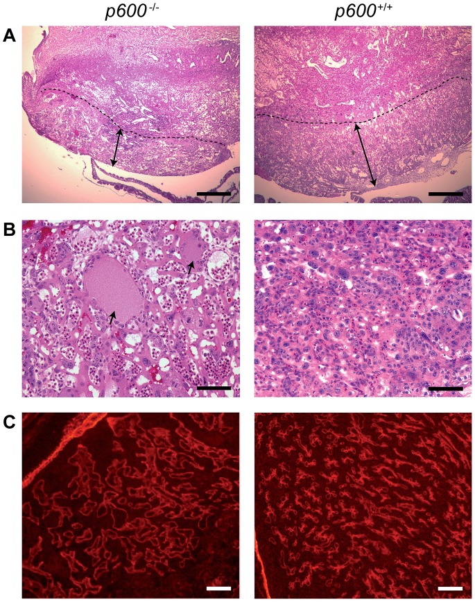
Figure 2. Placental abnormalities in p600 −/− animals.
(A) Placental labyrinth defects in p600 −/− animals. H&E staining images of the labyrinth layer of p600 knockout and control littermate at E12.5 (the area below the dashed lines) are shown. The labyrinth areas of p600 −/− placenta are thinner than those of wild type littermate as shown by arrows. Scale bars indicate 500 µm. (B) High magnification images of the labyrinth layer. Dilated blood vessels (arrows) are observed in p600 −/− placenta. Scale bars indicate 50 µm. (C) Immunofluorescence staining of blood vessels in labyrinth areas with anti-laminin antibody. The blood vessels in labyrinth are dilated and sparse in p600 KO animals at E12.5. Scale bars indicate 100 µm.

