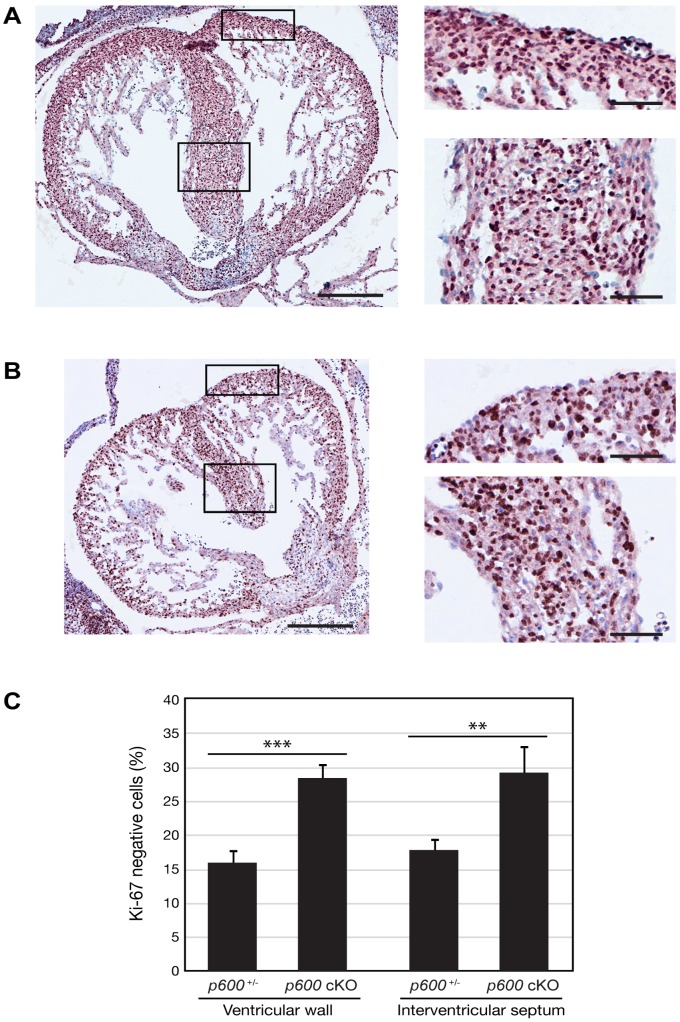
Figure 5. Proliferation defects in conditional p600 knockout heart.
Transverse sections of p600 −/+ (p600 +/−/ Sox2-Cre +) (A) and p600 cKO (B) heart at days E13.5 were stained with anti-Ki-67 antibody. After immunehistological staining, the samples were counterstained with hematoxylin. Magnified images of the ventricular wall (top) and the interventricular septum (bottom), the region indicated by rectangles, are shown in the right panels. Scales bars in the left and right panels indicate 500 and 200 µm, respectively. (C) The percentage of Ki-67 negative cells in the ventricular wall and the interventricular septum are shown. The p-values for Student’s t-test are shown in the graph. ** and *** show p-value <0.01 and <0.001, respectively.

