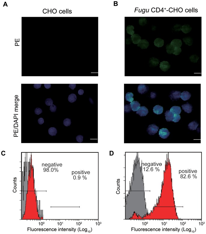
Figure 2. Reactivity of anti-Fugu CD4 Ab to Fugu CD4+-CHO cells.
Immunofluorescence of A) control CHO cells and B) Fugu CD4+-CHO cells. Cells were incubated with anti-Fugu CD4 Ab as primary Ab and goat anti-rabbit IgG (PE) as secondary Ab and DAPI to mark cell nuclei. Scale bar equals 20 µm. Stained cells C) control CHO cells and D) Fugu CD4+-CHO cells were also analyzed by flow cytometry. The setting of negative gate was used with the only secondary antibody reaction to the cells analysis (gray peak).

