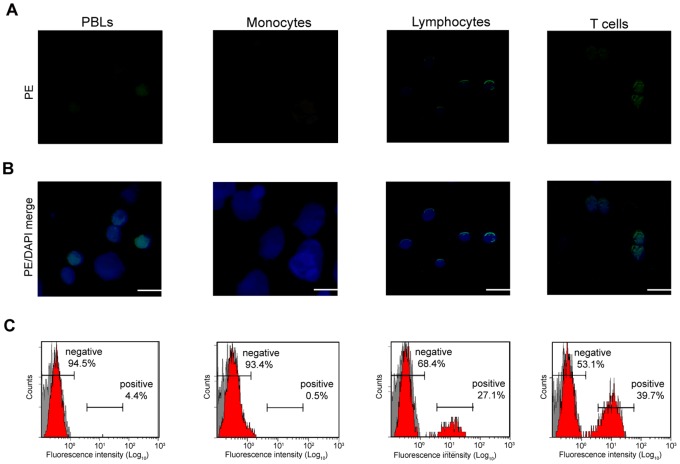
Figure 4. Immunofluorescence stains of Fugu CD4+ cells in PBLs, monocytes, lymphocytes and T cells.
A), B) Cells were reacted with anti-Fugu CD4 Ab as primary Ab and goat anti-rabbit IgG (PE) as secondary Ab and DAPI to mark cell nuclei. Scale bar equals 20 µm. C) Cells stained with anti-Fugu CD4 Ab were also analyzed by flow cytometry.

