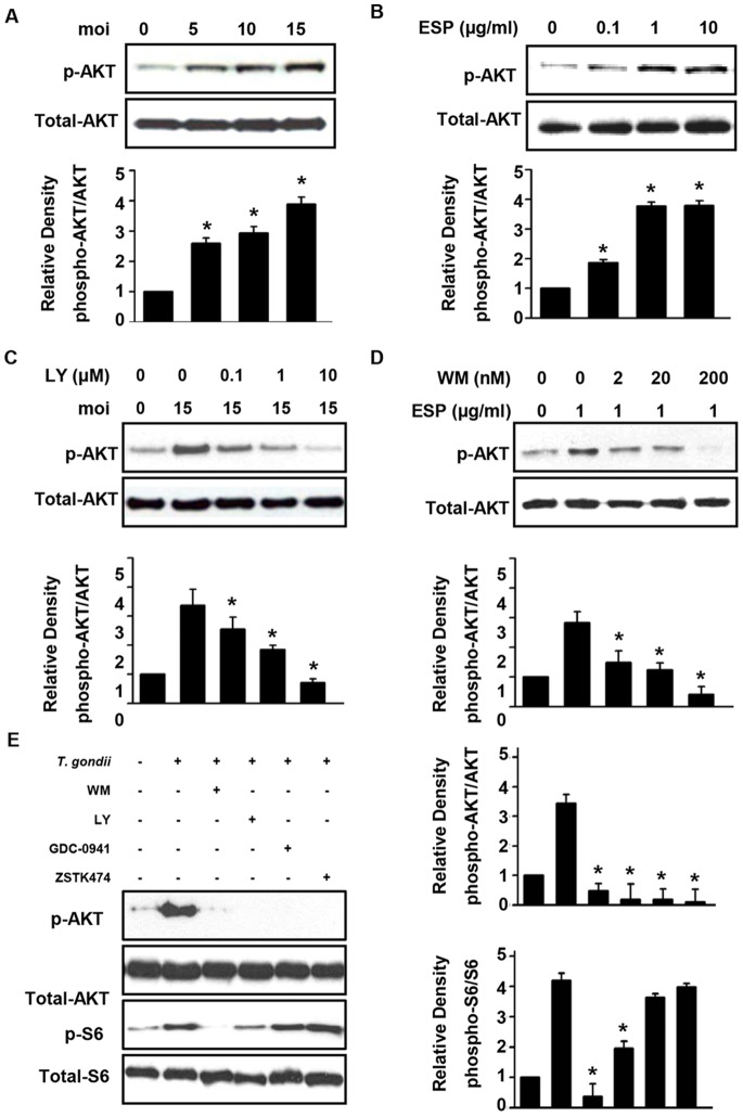
Figure 1. T. gondii and ESP activate the PI3K/Akt pathway in ARPE-19 cells.
ARPE-19 cells were deprived of serum for 1 day before use in subsequent experiments. (A) Cells were infected with GFP-RH tachyzoites at the indicated moi for 24 h. Two hours after infection, the extracellular tachyzoites were immediately removed by washing with PBS and then incubated in the absence of serum media for 22 h. (B) Cells were stimulated for 1 h with various concentrations of GFP-RH tachyzoites ESP ranging from 0.1 to 10 µg/ml. (C) ARPE-19 cells were infected with T. gondii (moi 15) for 24 h and treated with increasing concentrations of LY294002 for the final 1 h. (D) Cells were pretreated with wortmannin for 1 h, and then after washing, cells were stimulated with 1 µg/ml ESP for 1 h. Whole cell lysates were collected and analyzed by western blot using primary antibodies against phospho-Akt and total Akt. Akt phosphorylation levels were calculated as the ratio of untreated control cells after normalization to the amount of total Akt. (E) Cells were infected at moi of 15 for 23 h and then treated with either inhibitor for 1 h. Values represent the mean ± SD values of triplicates. Data are representative of three independent experiments. * denotes p<0.05 and indicates that differences were considered significant.

