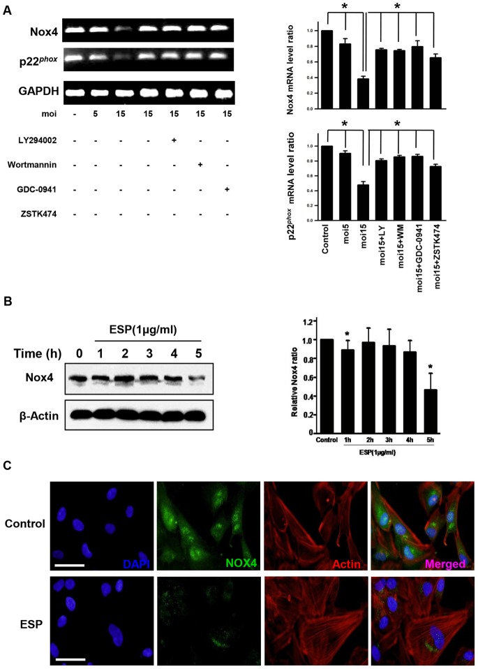
Figure 5. T. gondii and ESP suppressed host Nox4 expression via the PI3K/Akt pathway.
(A) RT-PCR analysis of the host Nox4 and P22phox mRNA levels in ARPE-19 cells. GAPDH was used as an internal control. (B) Cells were stimulated with 1 µg/ml ESP for the indicated times. Nox4 protein levels were analyzed by western blot, and β-actin was used as a loading control. (C) Immunocytochemistry of Nox4 in the ESP-stimulated cells. ARPE-19 cells were stimulated with 1 µg/ml ESP for 5 h. Cells were subsequently stained with anti-Nox4 antibody (green) and Texas Red®-X phalloidin for labeling F-actin (red). Nuclei were stained with DAPI (blue). The results are expressed as mean ± SD of three independent experiments. Scale bar = 100 µM. * denotes p<0.05, and these differences were considered significant.

