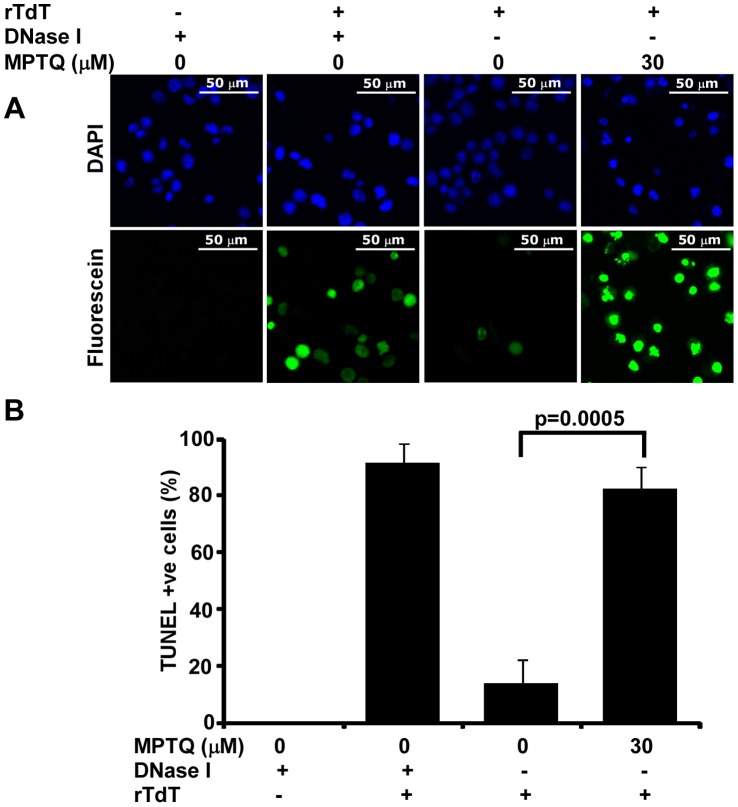
Figure 5. MPTQ induced nuclear DNA breaks in neuro 2a cells are positive for TUNEL staining, an indicator of apoptosis.
Neuro 2a cells were cultured and treated with 30 µM of MPTQ for 48 hrs. Control cells were treated with equal amount of DMSO. A) Fluorescent images of DAPI and fluorescein-12-dUTP were captured from multiple random fields using multi-dimension acquisition module of MetaMorph using identical settings and images were displayed with equal pixel intensity. Images display increased TUNEL positive neuro 2a cells in MPTQ treated cells than control cells B) Number of TUNEL positive cells were calculated using multi-cell scoring module of MetaMorph software and mean of three independent experiments are presented as histograms with standard deviation as error bars. Results indicated more than 80% cells were positive for TUNEL in MPTQ treated cells where as only 13% in control cells. p value was calculated by Student’s t-test and displayed. p≤0.05 is considered statistically significant.

