
Figure 7. MPTQ-mediated cell death is associated with increased phosphorylation of p53 at Ser15.
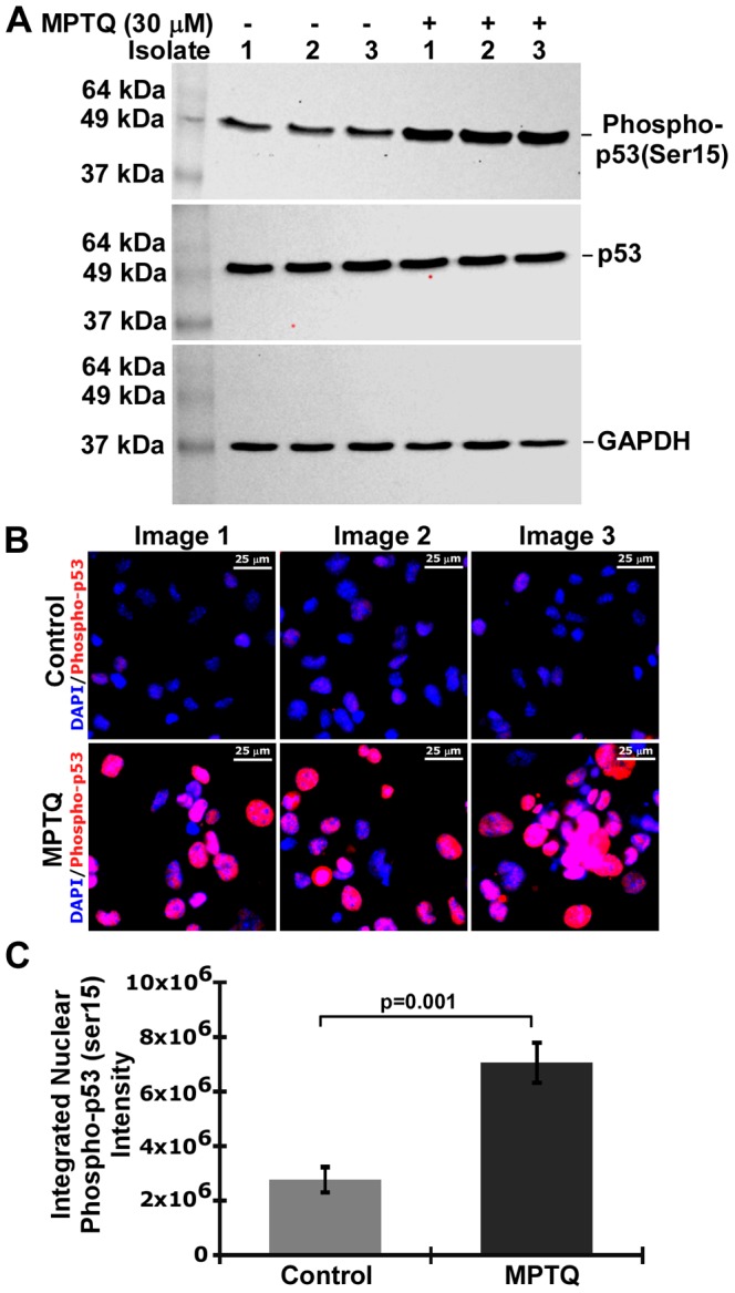
A) Western blot analysis of phospho-p53 (Ser15), p53 and GAPDH. Neuro 2a cells were either treated with 30 µM of MPTQ or DMSO alone for 24 hours. Three independent isolates were obtained and 60 µg of total proteins were size fractionated in 12% SDS-PAGE and western blotted either with anti-phospho-p53 (ser15) or with anti-p53 antibody. The blots were stripped and hybridized with anti-GAPDH antibody to normalize any loading difference. B) Immunocytochemistry of phopho-p53 (Ser15). Images represent three independent experiments C) Nuclear phospho-p53 (Ser15) intensity was measured as described in figure 6. Histograms represent mean integrated nuclear phopho-p53 (Ser15) intensity±SD of three independent experiments. p value calculated by Student’s t-test is displayed which indicates significant increased phosphorylation of p53 at Ser15 in MPTQ treated neuroblastoma cells.