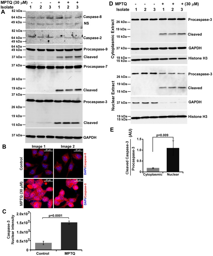
Figure 9. MPTQ-mediated cell death is associated with activation of caspases of intrinsic apoptosis pathway but not of extrinsic pathway.
A) Neuro 2a cells were cultured and treated with 30 µM of MPTQ for 24 hours and lysates were prepared. 60 µg of total proteins were resolved in 12% SDS-PAGE and immunoblotted with anti-caspase-8 or anti-caspase-2 or anti-caspase-9 or anti-caspase-3 or anti-caspase-7 antibody. Blots were stripped and immunoblotted with anti-GAPDH antibody. The results clearly indicate the activation of caspase-9, -3 and-7 but not caspase-8 and -2 in MPTQ treated cells. B) Immunocytochemistry of caspase-3 protein was performed as described earlier. Increased caspase-3 level was observed in the nucleus of MPTQ treated neuro 2a cells but not in control cells. C) Nuclear level of caspase-3 immunosignal was obtained using multi-cell scoring module and mean of three random images of two independent experiments were displayed as histograms. Error bar indicates standard deviation. D) Western blot analysis of cleaved caspase-3 level in cytosolic and nuclear fraction of MPTQ treated or untreated neuro 2a cells. Blots were also immunoblotted with anti-GAPDH and anti-histone H3 antibodies for normalization. E) Densitometric analysis of procaspase-3 and cleaved caspase-3 bands were made from cytosolic as well as from nuclear fractions. Cleaved caspase-3 to procaspase-3 ratio was obtained. Mean and standard deviation from three independent isolates were obtained and plotted as histograms. p value was calculated by Student’s t-test and is displayed which indicates significant increased mobilization of cleaved caspase-3 from cytoplasm to nucleus in MPTQ treated neuro 2a cells.

