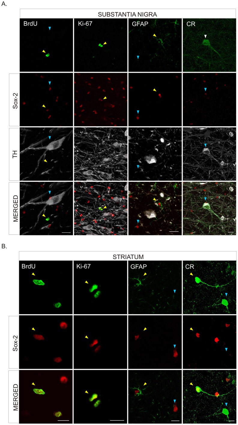
Figure 3. Characterization of Sox-2+ cells in the substantia nigra and striatum of naïve adult primates.
A. Confocal images show BrdU incorporation (yellow arrowhead), and co-expression of Ki67 (yellow arrowhead) in Sox-2+ cells in the parenchyma of the SNpc. Sox-2+ cells were never positive for TH. Some Sox-2+ cells corresponded to satellite glial cells sitting on TH+ neurons, as shown in the merged panels (blue arrowheads). Many Sox-2+ cells expressed GFAP (yellow arrowhead) but there were also Sox-2+/GFAP– (blue arrowhead) that may correspond to amplifying progenitors. There was no Sox-2+/CR+ co-expression in any cell in this region. Scale bar = 20 µm. B. In the striatal parenchyma some Sox2+ cell showed BrdU incorporation (yellow arrowhead) and co-expressed Ki67 (yellow arrowhead). Most Sox-2+ striatal cells expressed GFAP (yellow arrowhead) but there were also Sox-2+/GFAP– (blue arrowhead). Some CR+ striatal neurons showed nuclear expression of Sox-2 (yellow arrowhead) while others did not (blue arrowhead). Scale bars = 10 µm. Abbreviations: Substantia nigra pars compacta: SNpc; tyrosine hydroxylase: TH; glial fibrillary acidic protein: GFAP; calretinin: CR.

