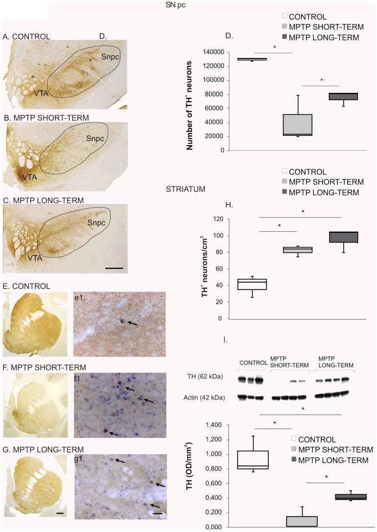
Figure 6. MPTP effect on the nigrostriatal system.
Midbrain sections at the level of the 3rd nerve from a control (A), a MPTP short-term (B) and a MPTP long-term treated monkey (C) immunostained for TH. Note the severe reduction of TH immunoreactivity in the SNpc of the lesioned monkeys. Scale bar = 1 cm. D. Stereological quantification of TH+ neurons in the SNpc. Data represent median and quartiles, N = 3, *p≤0.05. Images of precommisural striatum sections from a control monkey (E), a MPTP short-term monkey (F) and a MPTP long-term monkey (G) immunostained for TH. Note the loss of TH+ fibers in the lesioned monkeys. Scale bar = 1 cm. Representative striatal sections of a control (e’) a MPTP short-term monkey (f’) and a MPTP long-term monkey (g’), at higher magnification showing the intrinsic TH+ neurons (arrows). Scale bar = 300 µm. H. Quantification of striatal TH+ neurons. There was a significant increase in the density of TH+ neurons after MPTP administration in both groups. Data represent median and quartiles, N = 3, *p≤0.05. I. Western blot to assess striatal TH levels showing a reduction in the MPTP-short term group and a partial recovery in the MPTP-long term group with respect to controls. Data represent median and quartiles, N = 3, *p≤0.05. Abbreviations: 1-methyl-4-phenyl-1,2,3,6 tetrahydropyridine: MPTP; tyrosine hydroxylase: TH; substantia nigra pars compacta: SNpc.

