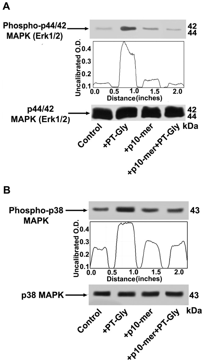
Figure 2. Inhibitory effect of gliadin peptide 10-mer on ERK and p38 MAPK phosphorylation.

CACO-2/TC7 cells either unstimulated or stimulated with PT-Gly (1mg/ml), p10-mer (50 µg/ml), p10-mer (50 µg/ml) + PT-Gly (1mg/ml), were analyzed by Western blot for ERK and p38 phosphorylation. Phosphorylated levels of ERK were analyzed in whole cell extracts by Western blot with anti-phospho-ERK1/2 antibodies; for control, the blotted membranes were stripped and reprobed with anti-ERK1/2 antibodies. Bound antibodies were visualized with HRP-conjugated IgG and immunoreactivity was assessed by ECL. (B) Phosphorylated levels of p38 MAPK were analyzed in whole cell extracts by Western blot with anti-phospho-p38 MAPK antibodies; for control, the blotted membranes were stripped and reprobed with anti-p38 MAPK antibodies. Bound antibodies were visualized with HRP-conjugated IgG and immunoreactivity was assessed by ECL. Densitometric analysis was performed using ImageJ version 1.46 software and peaks were reproduced by reading the Western Blot bands. One example representative of 3 experiments.
