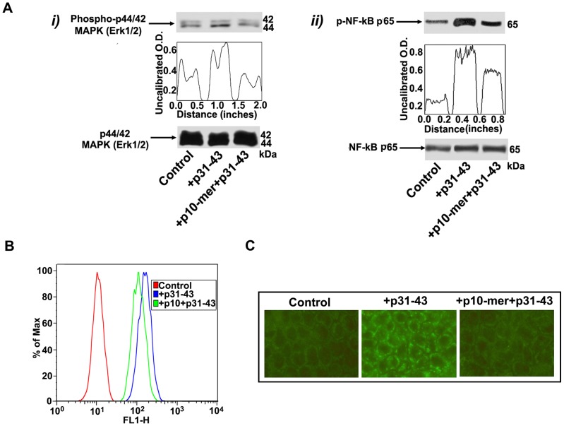
Figure 5. Effect of gliadin peptide 10-mer on p31–43 activity and entrance in CACO-2 cells.
(A) CACO-2/TC7 cells, either unstimulated or stimulated with p31–43 (50 µg/ml), p10-mer (50 µg/ml) + p31–43 (50 µg/ml), were analyzed by Western blot for ERK phosphorylation and NF-kB activation. (i) Phosphorylated levels of ERK were analyzed in whole cell extracts by Western blot with anti-phospho-ERK1/2 antibodies; for control, the blotted membranes were stripped and reprobed with anti-ERK1/2 antibodies. Bound antibodies were visualized with HRP-conjugated IgG and immunoreactivity was assessed by ECL. (ii) NF-kB activation was analyzed in whole cell extracts by Western blot with anti-phospho-NF-kB p65 Ser antibodies; for control, the blotted membranes were stripped and reprobed with anti-NF-kB p65 antibodies. Bound antibodies were visualized with HRP-conjugated IgG and immunoreactivity was assessed by ECL. Densitometric analysis was performed using ImageJ version 1.46 software, peaks were reproduced by reading the Western Blot bands. One example representative of 3 experiments. (B) CACO-2/TC7 cells, either unstimulated or stimulated with biotinylated p31–43 (50 µg/ml), p10-mer (50 µg/ml) + biotinylated p31–43 (50 µg/ml), were immunostained with streptavidin-AlexaFluor and analyzed by a BD FACSCalibur flow cytometer. (C) CACO-2/TC7 cells, either unstimulated or stimulated with biotinylated p31–43 (50 µg/ml), p10-mer (50 µg/ml) + biotinylated p31–43 (50 µg/ml), were immunostained with streptavidin-AlexaFluor. The images were acquired using an Olympus U RFL fluorescence microscope.

