
Figure 2. Resting state functional connectivity of the default mode.
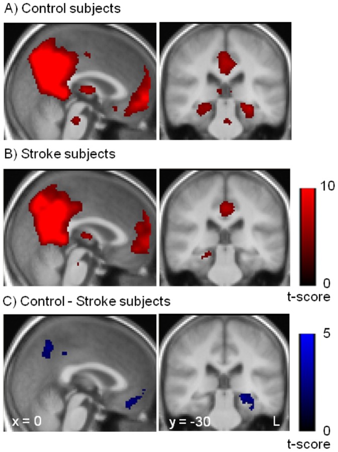
Spatial maps of the default mode network for control subjects (A) and for stroke subjects (B). (C) Resting state functional connectivity differences between control and stroke subjects. Stroke subjects showed decreased functional connectivity in the posterior cingulate gyrus, medial prefrontal cortex and left medial temporal lobe. The statistical maps are superimposed onto the spatially normalized and averaged group T1-images.