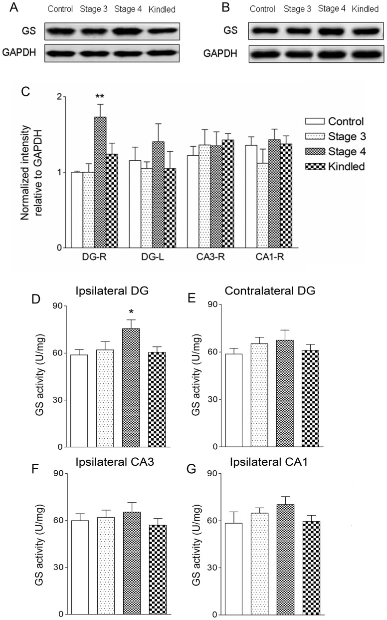
Figure 2. Upregulation of GS function in the ipsilateral DG in the rat amygdala kindling model.
Measurements of GS expression in the ipsilateral DG (A) and CA3 (B) by western blotting and enzyme activity in bilateral DG (D and E), ipsilateral CA3(F) and CA1(G) in the progression of amygdala kindling (control group, n = 6/stage; stages 1–4 and fully kindled, n = 5–7/stage). Data are shown as mean ± SEM. * P<0.05 and **P<0.01 compared with controls.

