
Figure 11. Western blotting of virion and cell lysate samples collected from cells transfected under various conditions.
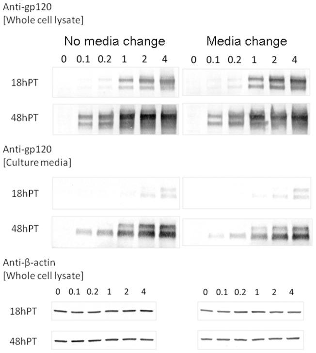
The amount of envelope plasmid in μg is indicated above the lanes. The double bands from anti-gp120 staining corresponded to gp120 (bottom) and gp160 (top), respectively. β-actin expression was also probed simultaneously to validate the comparison across different transfection conditions. Two independent analyses yielded results that support the same conclusions.