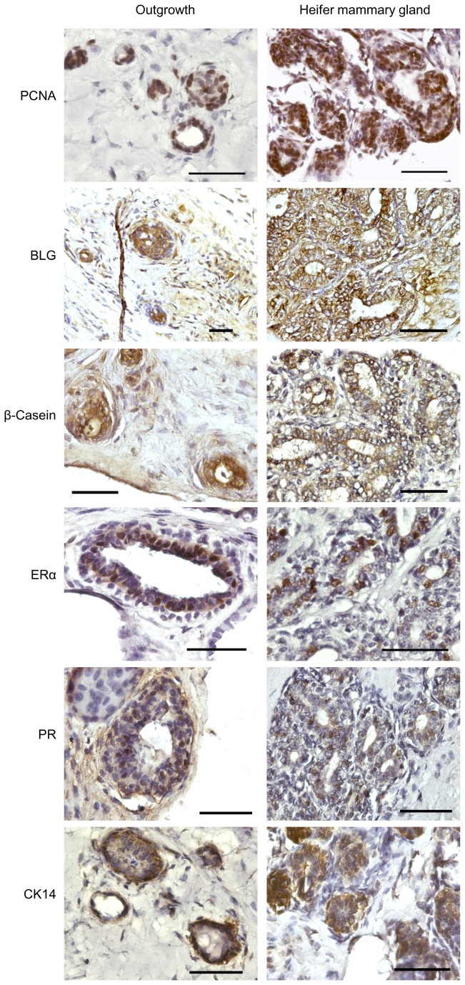
Figure 2. Bovine mammary outgrowths developed in the mouse, resemble the immunohistochemical characteristics of the donor tissue.
Immunohistochemical analyses of selected markers on paraffin sections from bovine outgrowths (left panels) and mammary epithelium from heifer’s mammary gland (right panels). The outgrowths and the heifer’s tissue were subjected to similar histological procedures, including Carmine staining, prior to immunohistochemistry and hematoxylin counterstaining. Bar = 50 µm.

