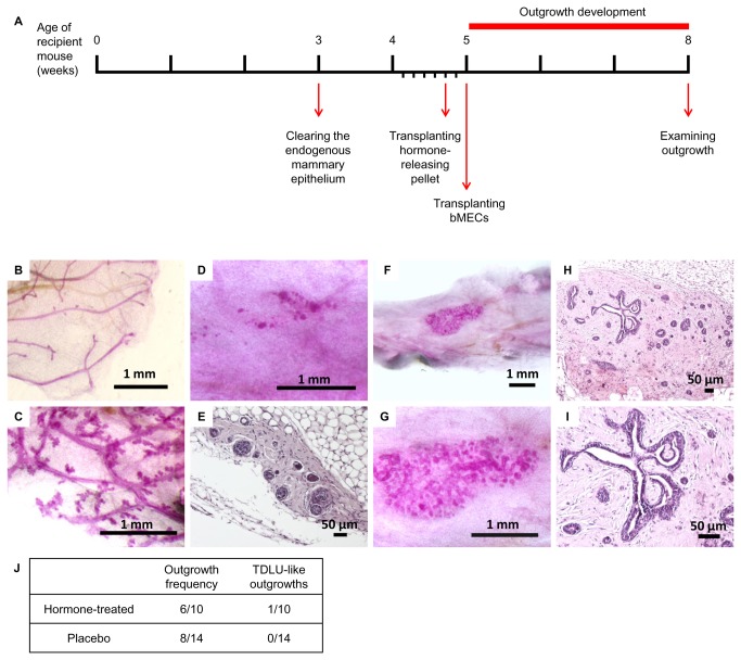
Figure 3. A TDLU-like structure develops from transplanted bovine cells in mice treated with estrogen and progesterone.
A: Timeline of hormone treatment and bMEC transplantation. Hormone pellets were inserted 2 days prior to bMEC transplantation. B, C: Carmine-stained endogenous mammary epithelium from recipient mice carrying placebo (B) or hormone pellets (C). Enhanced branching of the endogenous ductal network is noted in (C). D, E: Carmine-stained wholemount (D) and H&E-stained paraffin section (E) depicting typical morphology of bovine outgrowth in the cleared mammary fat pad of hormone-treated mice. F–I: Carmine staining of mammary wholemounts (F, G) and H&E staining of paraffin sections (H, I), depicting a particular case of an outgrowth exhibiting significant growth and TDLU-like morphology under continuous systemic treatment of estrogen and progesterone. J: Frequency and morphology of outgrowths developed from transplanted bMECs in the cleared mammary fat pad of recipient mice that received, or did not receive, hormone treatment.

