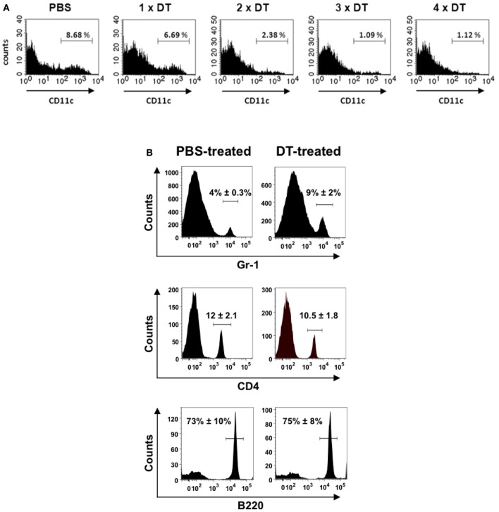
Figure 1.
Depletion of CD11c+ cells in the lungs of CD11c-DTR chimera mice after treatment with successive doses of DT. CD11c-DTR mice were injected with 1, 2, 3, or 4 successive doses of DT (8 ng/g body weight) or PBS. Lungs were digested and transformed in single cell suspensions 24 h after treatment and stained with PE-conjugated anti-CD11c antibodies for FACS analysis. (A) Representative histogram of CD11c expression in lung cells of PBS-treated or DT-treated CD11c-DTR mice. (B) Percentage of neutrophils (Gr-1+) (upper histograms), CD4+ lymphocytes (middle histograms) and B cells (B220+) (lower histograms) in the lungs of PBS-treated (left) or DT-treated (right) CD11c-DTR mice. The mean ± SD percentages of positive cells are indicated in each histogram. Data shown are representative of one out of three separate experiments.

