Figure 7.

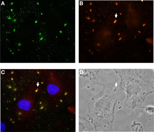
Poor uptake of S. pneumoniae by bone marrow-derived DCs in in vitro assays. Bone marrow-derived DCs were infected with S. pneumoniae for 2 h, fixed, and stained for with polyclonal rabbit anti-S. pneumoniae antibodies, followed by Alexa green-conjugated goat anti-rabbit antibodies (A). Cells were then permeabilized with 0.025% Triton X-100 in PBS, and intracellular bacteria were stained by anti-S. pneumoniae antibodies, followed by Alexa red-conjugated goat anti-rabbit antibodies (B). In the merged image shown in (C), extracellular bacteria are yellow-green, intracellular bacteria are red and the DNA in the nucleus of DCs is stained in blue. The white arrow indicated an intracellular S. pneumoniae. (D) Phase contrast picture showing the contour of the DCs.
