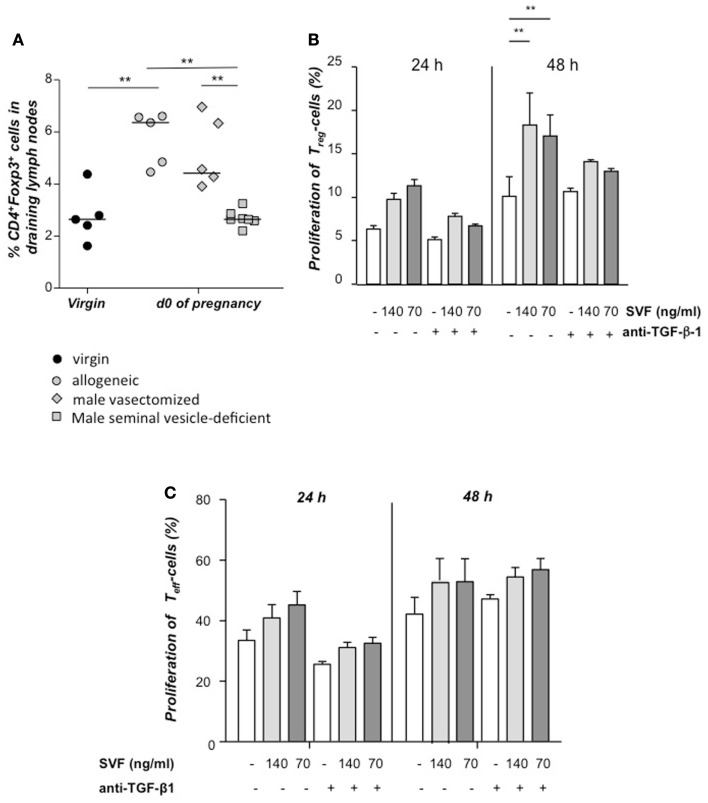
Figure 2.
Tregs expand in vivo and in vitro in presence of seminal fluid. Percentage of CD4+Foxp3+ cells in the uterine draining lymph nodes of CBA/J females mated with seminal vesicle-deficient, vasectomized, or intact BALB/c males was analyzed at day of conception (day 0.5) and compared to virgin CBA/J females (n = 4–7) (A). Statistical analysis were performed by Mann–Whitney-U test (**P ≤ 0.01). (B) Tregs were isolated from non-pregnant CBA/J females by magnetic cell sorting, stained with CFSE and cultured with seminal vesicle fluid (SVF) from BALB/c males. TGF-β1 was blocked with anti- TGF-β1 antibody, and proliferation of Treg determined after 24 h by using FACScan Calibur. Data are representative of four experiments and expressed as mean with SEM. Analysis was performed by two-way ANOVA test (**P ≤ 0.01). (C) Conventional T effector cells were isolated from non-pregnant CBA/J females by magnetic cell sorting, stained with CFSE and cultured with seminal vesicle fluid (SVF) from BALB/c males. TGF-β1 was blocked with anti- TGF-β1 antibody, and proliferation of Treg determined after 24 h by using FACScan Calibur. Data are representative of four experiments and expressed as mean with SEM. Analysis was performed by two-way ANOVA test and no statistically significant differences were found among the groups.

