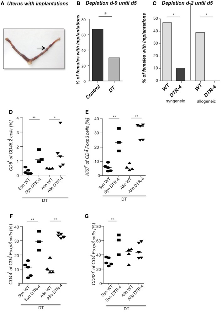
Figure 3.
Embryo implantation is impaired after Treg depletion. Foxp3+ Treg were depleted in Foxp3DTR mice by application of DT every fourth day, starting 9 days before mating (day 9) with BALB/c males. Control groups received PBS. (A) shows a representative picture of a uterus stained at day 5 post conception with Chicago Blue dye application. The arrow indicates a representative implantation site. The percentage of females presenting implantations was analyzed on day 5 after mating [(B), n = 5–12]. Foxp3+ Treg were depleted in Foxp3.LuciDTR-4 mice by daily application of DT, starting on day 2 with allogeneic (CBAJ) or syngeneic (C57/BL6) males. Control groups were wt C57/BL6 females treated with DT. Implantation numbers were measured at day 5 [(C), n = 15–21]. For (B,C), data are expressed as medians of % of implanted females and analyzed by Fisher’s exact test (#P < 0.1, *P < 0.05). Not all plugged animals became pregnant. In samples from animals shown in Figure 3B, the percentage of CD8+ cells was analyzed in the uterus by flow cytometry (D), and the percentage of KI67+, CD44+, and CD62L+ (E–G) determined in uterine draining lymph nodes (n = 5/group). Data are expressed as medians and were analyzed by Mann–Whitney-U test (*P < 0.05; **P < 0.01).

