Figure 6.
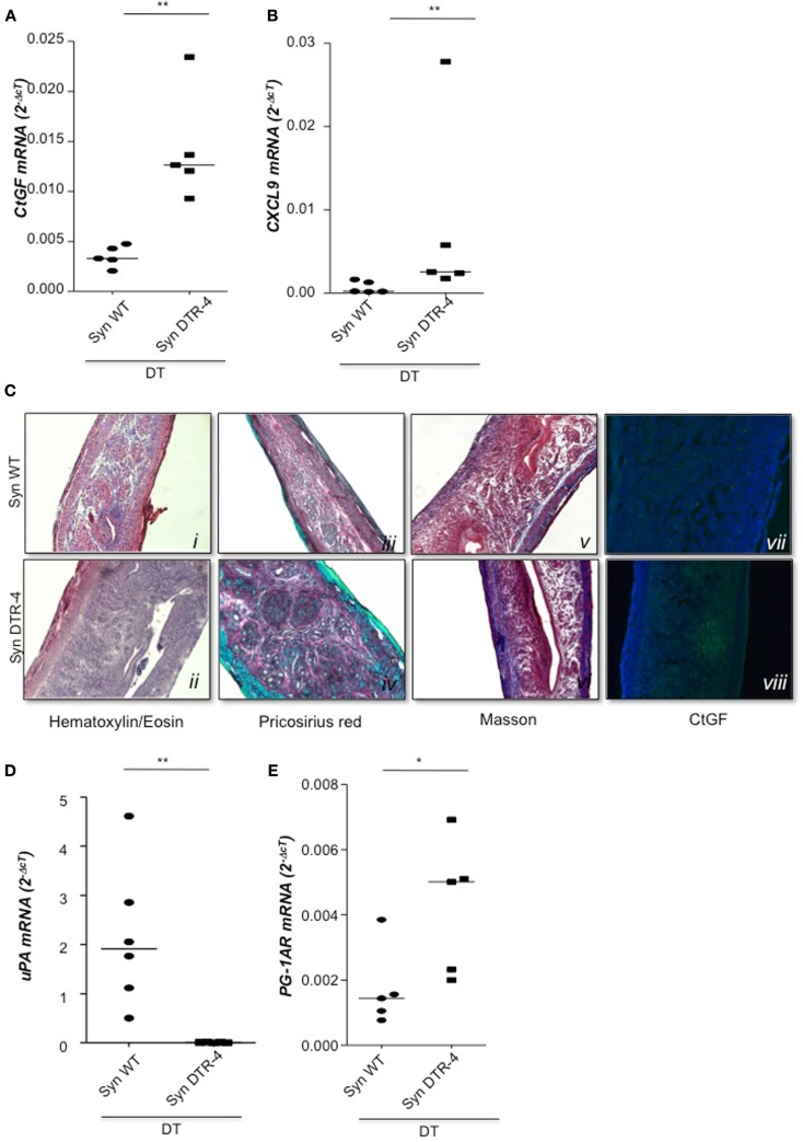
Treg depletion leads to uterine fibrosis. In samples from Foxp3.LuciDTR-4 or wild type animals treated with DT by daily application starting on day 2 and further mated syngeneically, fibrosis markers were measured (n = 4–6/group) in uterine tissue by qPCR. Levels of CtGF (A) and CXCL9 (B) were significantly elevated in mice depleted of Tregs as compared to DT-treated controls. Data are expressed as single dots with medians and were analyzed by Mann–Whitney-U test (#P < 0.1, **P < 0.01). Additionally, staining with Hematoxylin/Eosin revealed fibrosis areas with disorganized collagen fibers in animals without Tregs compared to wild type controls (C ii vs. i). Pricosirius red staining revealed a higher amount of green/blue-stained thin collagen fibers that confirms fibrosis in mice devoid of Tregs vs. controls (C iv vs. iii). Further, Masson’s staining manifested larger areas of collagen fibers in blue/violet in DT-treated Foxp3.Luci.DTR animals compared to DT-treated controls (C vi vs. v). Finally in (C vii, viii), immunofluorescence staining for CtGF confirmed accumulation of CtGF positive cells in mice depleted in Tregs as compared to the controls. All pictures were taken with a 10× objective. Analysis of uPA mRNA revealed that animals depleted in Tregs presented very low, almost undetectable levels of this molecule as compared to DT-treated controls (D). Treg depletion further provoked an augmentation of prostaglandin 1A mRNA (E).

