Figure 4.
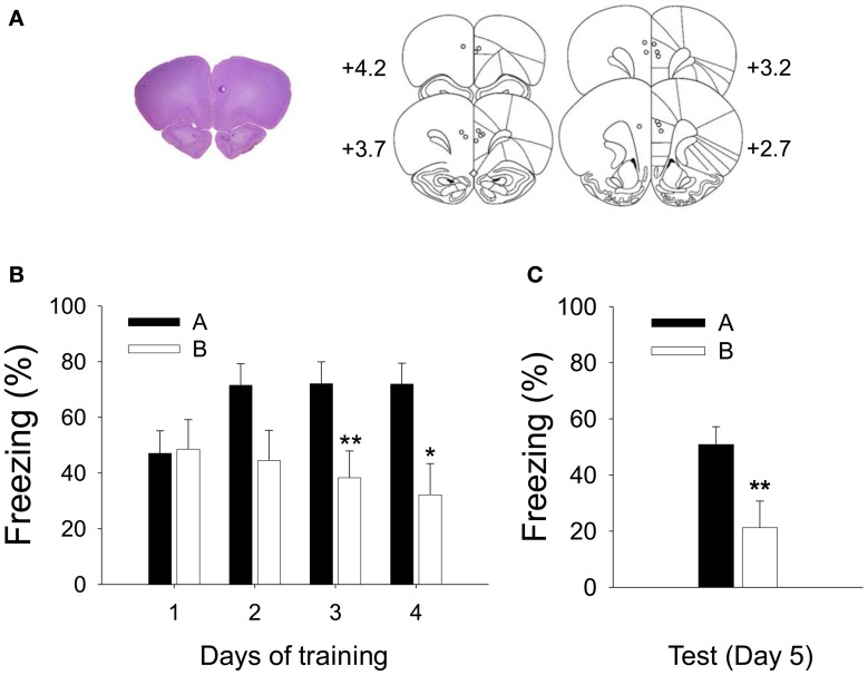
Recorded locations and acquisition of CDFD. (A) A high-resolution scan of a cresyl-violet-stained coronal section shows a representative electrode placement in the PL marked by a small lesion (left), and the reconstruction of electrode placements from all subjects (right) shows that the recording locations were confined within the PL. (B) Robust CDFD was developed following 4 days of training as all animals exhibited significantly greater freezing to the CS in Context A than in Context B on days 3 and 4. (C) On the test session, they also showed significantly more freezing to the CS in Context A than in Context B. * and **denote p < 0.05 and p < 0.01, respectively.

