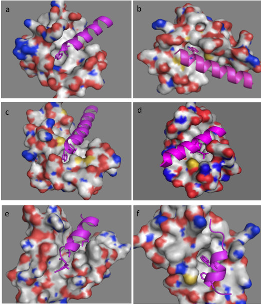
Figure 2.

3D structures of the complexes formed by: (a) human centrin 2 and a 10 residue peptide of Xeroderma Pigmentosum group C protein, code entry 2GGM. (b) chicken calmodulin and smooth muscle myosin light chain kinase (smMLCK), code entry 2O5G. (c) scherffelia dubia centrin and smMLCK peptide, code entry 3KF9. (d) rabbit cardiac troponin C and a fragment of cardiac troponin I, code entry 1A2X. (e) human BCL-XL and BAK peptide, code entry 1BXL. (f) human E3 ubiquitin-protein ligase MDM2 and p53 tumor transactivation domain, code entry 1YCR. All proteins are shown as surface in atom color type (C and H-white, N – blue, O -red, S – yellow) and ligands are shown in magenta cartoon with hydrophobic interacting residues given as sticks.
