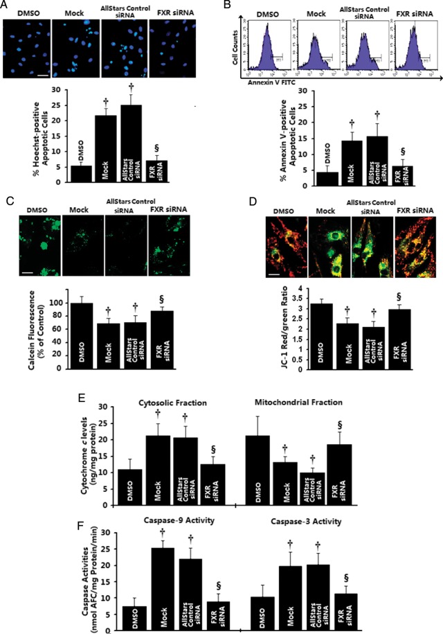
Figure 4.
Effects of in vitro farnesoid-X-receptor (FXR) silencing on apoptotic mitochondrial changes in cardiomyocytes. Neonatal rat ventricular myocyte (NRVMs) were transfected with 20 nmol/L of farnesoid-X-receptor siRNA, AllStars Negative siRNA, or mock-treated for 24 h, followed by GW4064 (5 μmol/L) treatment for 12 (B–F) or 24 h (A). DMSO-treated cells were utilized as control. (A and B) Late apoptosis assessed by Hoechst staining (A, n= 4 independent experiments; bar= 40 μm) and early apoptotic cells as assessed by annexin V staining (B, n= 3 independent experiments). (C) Representative confocal images of mitochondrial permeability transition pore opening measured by calcein-AM/CoCl2 quenching (upper) and quantitative data by flow cytometry (bottom, n= 3–4 independent experiments). (D) Representative confocal images of mitochondrial depolarization by JC-1 staining (upper) and quantitative data by flow cytometry (bottom, n= 3–4 independent experiments). (E) Mitochondrial and cytosolic cytochrome c levels measured by sandwich ELISA (n= 4 independent experiments). (F) Caspase-9-like and caspase-3-like activities were measured using the colorimetric substrate LEHD-AFC or DEVD-AFC (n= 3–4 independent experiments). *P< 0.05, †P< 0.01 compared with vehicle-treated cells, ‡P< 0.05, §P< 0.01 compared with GW4064-treated cells.

