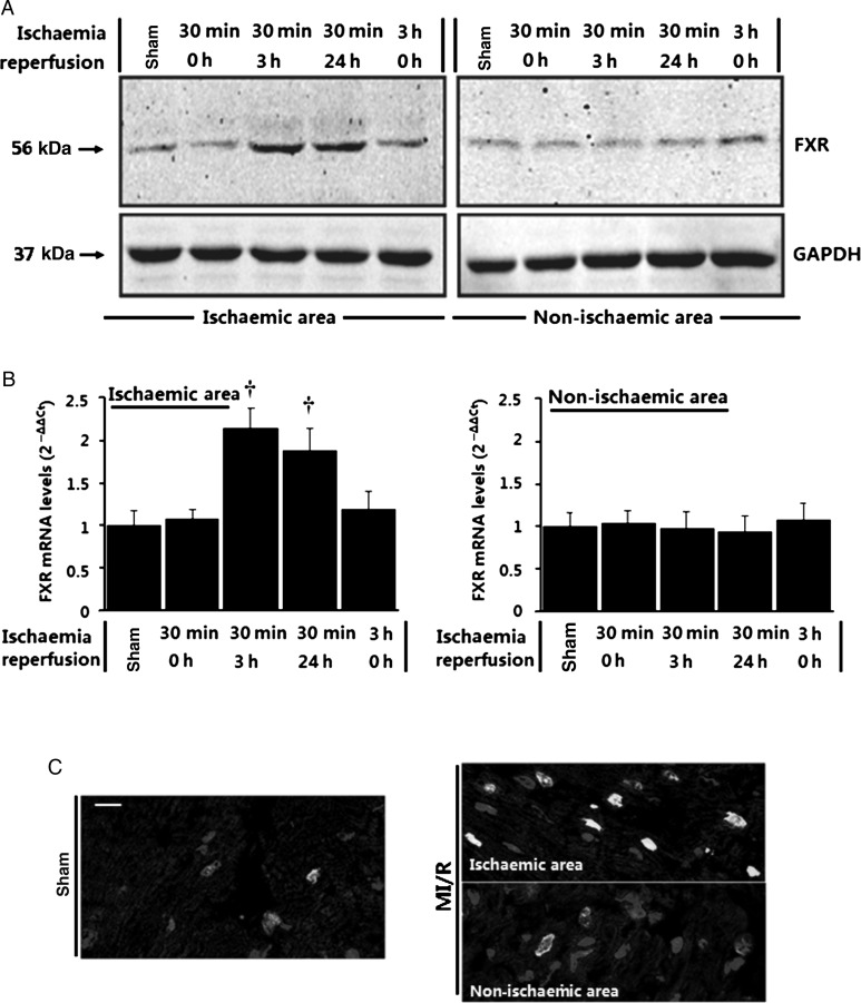
Figure 5.
Upregulation of farnesoid-X-receptor (FXR) by ischaemia/reperfusion stimuli in the heart. (A and B) Time course of farnesoid-X-receptor activation detected by western blots (A) and real-time quantitative PCR (B) in the heart (n= 5–6 per time point, ischaemic and non-ischaemic areas) subjected to in vivo ischaemia/reperfusion (I/R) for the indicated times. Sham-operated animals were utilized as control. Results were normalized against GAPDH and converted to fold induction relative to sham-operated controls. *P< 0.05, †P< 0.01 compared with sham-operated controls. (C) Confocal immunofluorescence analysis on heart sections from sham-operated and ischaemia/reperfusion mice 24 h after surgery. Sections were subjected to immunofluorescence for farnesoid-X-receptor (green) and α-actinin (red) and to staining for nuclei with DAPI (blue), and overlays are shown (Bar= 20 μm).

