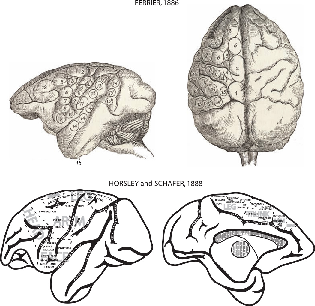
Fig. 2.
Montage depicting motor organization of the cerebral cortex determined by the application of electrophysiologic stimulation of the cortical surface in monkeys. Top: Lateral (left) and dorsal (right) views of the cortex with distinct movement representations outlined by irregular circles with numbers published by the British neurologist David Ferrier [29]. The sites that evoked movements in the upper limb are numbered 4 (“retraction with abduction of the opposite arm”), 5 (“extension forward of the opposite arm and hand”), a, b, c, d (“individual and combined movements of the fingers and wrist” for prehension of the opposite hand), 6 (supination and flexion of the forearm). Bottom: Lateral (left) and medial (right) views of the cortex with movement representations published by Horsley and Schafer [45]. This map provides one of the most comprehensive representations of the motor cortex published in the 1800s. Of notable significance was the early recognition of head, arm, trunk, and leg representations on the lateral surface of the hemisphere as well as the medial surface.

