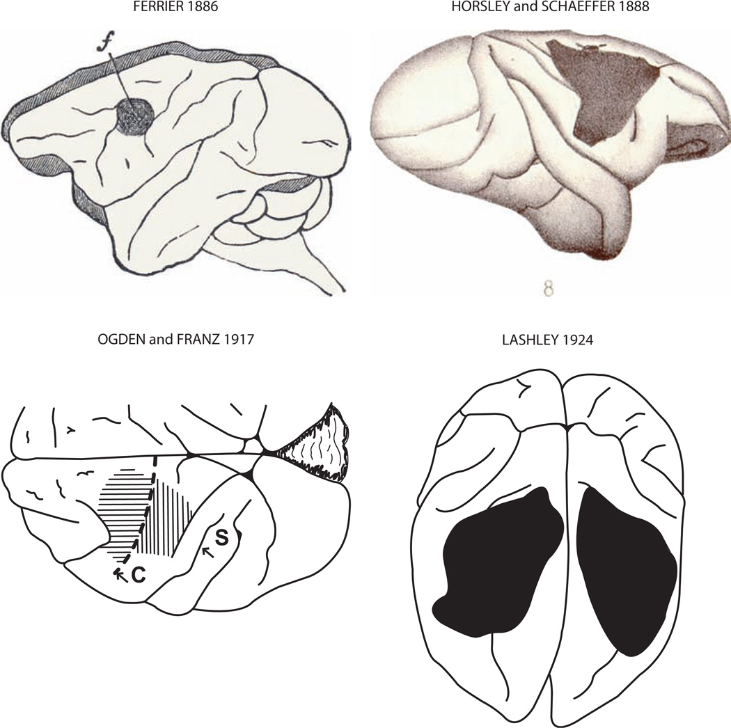
Fig. 3.
Montage depicting the precentral motor lesion site in monkeys in the classic studies of Ferrier [29], Horsley and Schaffer [45], Ogden and Franz [79] and Lashley [52] (Fig. 1, American Medical Association, Archives of Neurology and Psychiatry, reproduced with permission). In the Ogden and Franz map, the horizontal lines indicate the first surgical ablation which involved the excitable precentral motor cortex. The vertical hatching over S1 indicates an apparent abnormality of that area. In the other maps, the frontal motor lesion site is represented by the blackened region.

