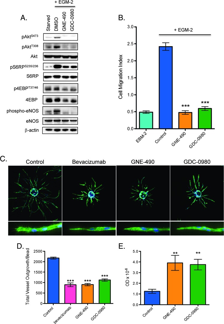
Figure 6.
Inhibition of PI3K reduces endothelial cell migration, sprout formation, and viability in vitro. (A) HUVECs were stimulated with EGM-2 growth factor media for 1 hour in the presence of DMSO vehicle (control), 0.40 µM GNE-490, or 0.40 µM GDC-0980 before immunoblot analysis for PI3K pathway markers and eNOS as shown. (B) HUVEC migration was induced with EGM-2 growth factor media in the presence of DMSO (control), 0.4 µM GNE-490, or 0.4 µM GDC-0980 for 72 hours and quantified as mean ± SEM. (C) Representative images of established endothelial sprouts after treatment with 20 µg/ml control antibody (anti-ragweed), 20 µg/ml bevacizumab, 0.40 µM GNE-490, and 0.40 µM GDC-0980 for 4 days. Cells were imaged using Molecular Devices ImageXpress Micro automated microscope with a 4x S Fluor objective and (D) quantified for total vessel outgrowth/bead (mean ± SEM). (C, bottom row) Higher resolution representative images (10x) of endothelial sprout tips. (E) Endothelial cell apoptosis was measured by a nuclear ELISA assay after treatment with DMSO vehicle (control), 0.40 µM GNE-490, or 0.40 µM GDC-0980 for 48 hours and quantified as mean optical density ± SEM. **P < .01, ***P < .001 compared to control using Dunnett's method.

