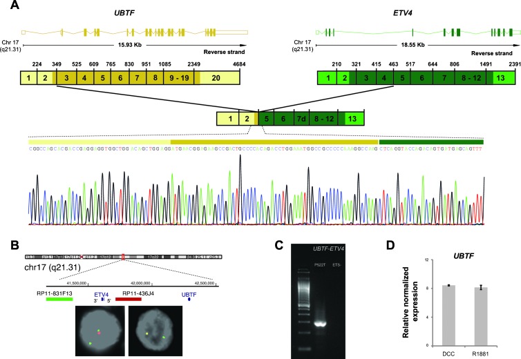
Figure 3.
Characterization of the gene fusion involving UBTF and ETV4 in the clinical prostate cancer P522T with ETV4 outlier expression and with an ETV4 genomic rearrangement previously demonstrated by FISH. (A) Fusion transcript of the untranslated exon 1 and exon 2 of UBTF with exon 5 of ETV4 detected by sequencing of the 5′RACE PCR products using a reverse primer on exon 10 of ETV4. The upper representation depicts wild-type transcripts of UBTF and ETV4 indicating their length and chromosomal localization. Exons are represented by boxes (colored if translated) and introns by horizontal lines. The scheme below represents the wild-type cDNA, where untranslated regions are in lighter shades. (B) Interphase FISH on formalin-fixed, paraffin-embedded tissue of the PCa P522T. Deletion of the probe spanning the chromosome region between UBTF and ETV4 (red) indicates a cryptic deletion at chromosome 17 as the genomic mechanism for UBTF-ETV4 gene fusion. (C) RT-PCR assay for detection of UBTF-ETV4 fusion transcript in the PCa P522T and in an ETS-negative PCa using a forward primer in exon 2 of UBTF and a reverse primer in exon 6 of ETV4. (D) Expression of UBTF is relatively unchanged in LNCaP cell lines treated with an androgen analog (R1881) versus cells grown in dextran-coated charcoal culture medium as a control. The expression data were obtained from the Gene Expression Omnibus (GSE32875).

