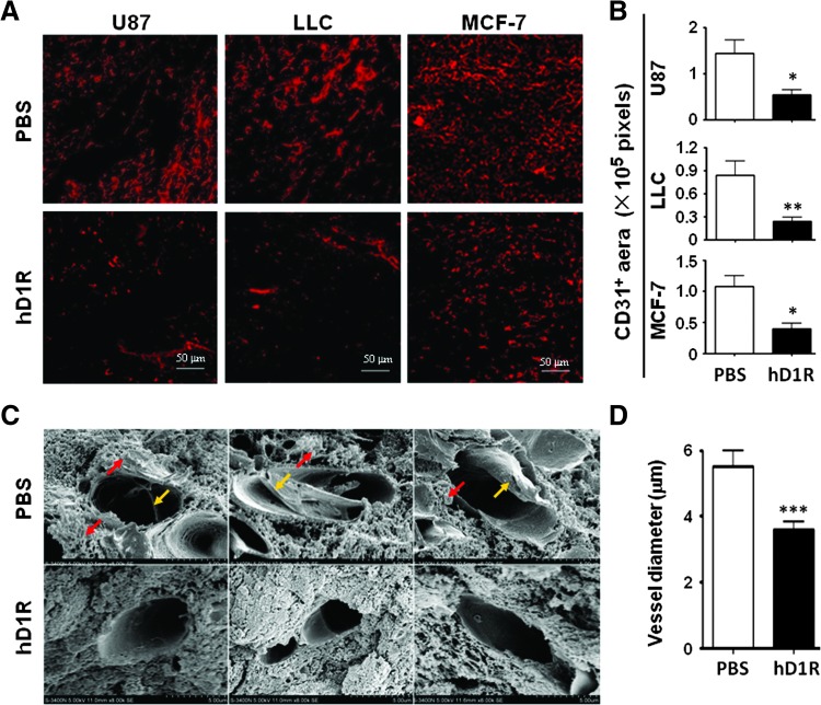
Figure 5.
hD1R reduced tumor vasculature. (A, B) U87, LLC, and MCF-7 cells were inoculated s.c. in nude mice. The mice were injected i.p. with PBS or hD1R twice a week from the 7th day of the tumor inoculation. Tumors were dissected on the last day of the experiments, sectioned, and immunostained with anti-CD31. CD31+ areas per field were quantified and were compared between groups (B). (C, D) U87 tumors in A were dissected, sectioned, and observed under SEM (original magnification, x 8000). The yellow arrows and red arrows indicate fibrous materials and leakage-like materials within and outside the microvessels. The shortest inside diameter of each vessel section (20 vessels for each group) was measured and compared (D). Bars, means ± SD. *P < .05, **P < .01, ***P < .001, n = 8.

