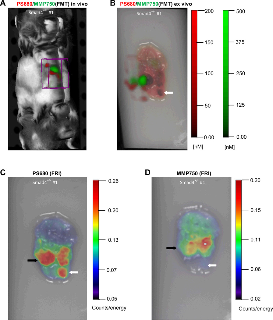
Figure 1.
In vivo and ex vivo imaging of gastric tumors in a Smad4+/− mice injected with activatable cathepsin and MMP probes using FMT and FRI. (A) In vivo FMT image of Smad4+/− mouse. ROI (boxed area) reveals NIRF at specific emission wavelengths for each probe in the anatomical region of the stomach. (B) Ex vivo FMT imaging of stomach from Smad4+/− mouse shows signal in visible gastric tumors. The 3D FMT image is superimposed on the 2D FRI image. (C) 2D FRI image of gastric tumor activated by cathepsin. (D) 2D FRI image of gastric tumors activated by MMP. Noted are differences between the two probes: the open arrow demonstrates probe activation with cathepsin but not MMP; and the closed arrow showing a neoplasm with cathepsin versus MMP.

