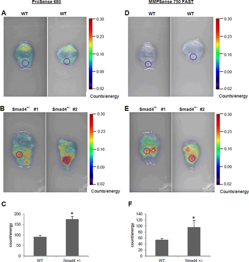
Figure 3.
Ex vivo FRI imaging of stomach tissue from WT and Smad4 +/− mice injected with cathepsin and MMP molecular probes. Stomach tissues were dissected immediately after in vivo imaging and imaged at 680 and 750 excitation wavelengths. Identical ROI (circles) were placed on area of interest in WT and Smad4+/− mice stomach. (A, D) Representative FRI images from WT. (B, E) Representative FRI images from Smad4+/− mice injected with the cathepsin and MMP probes (D). (C, F) 2D quantification of total counts (counts/energy) in WT and Smad4+/− mice. Histograms show mean ± SEM; n=4 for Smad4 +/− mice and n=3 for the WT mice; *p=<0.05 compared with WT.

