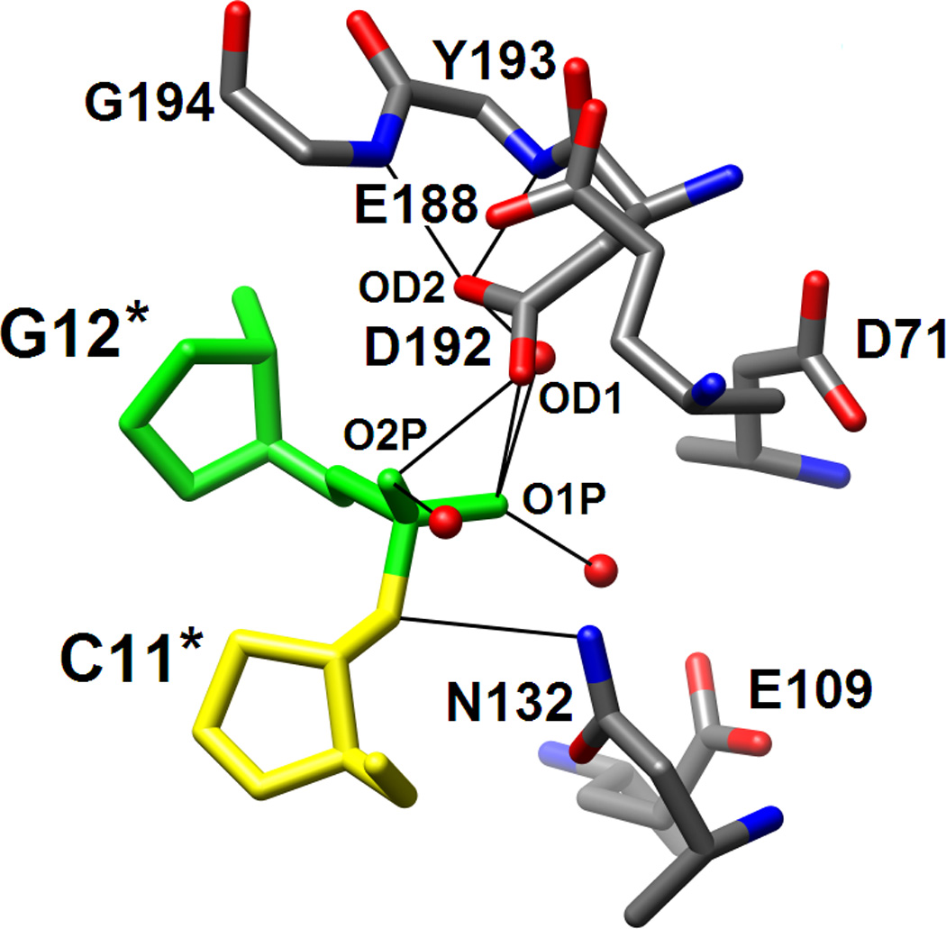
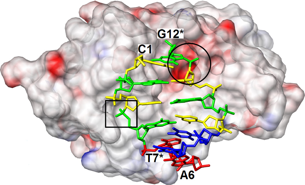
Figure 5.

RNase H active site location and configurations in the complexes with DDD and RNA/DNA hybrid. (A) Electrostatic surface potential of Bh-RNase H; red and blue regions indicate negative and positive charge, respectively, and the potential scale ranges from −50 to +50. Only half the DDD duplex is shown and the color code of nucleotides is identical to that in Fig. 1. The locations of the active site (circled) and the phosphate binding site (boxed) can easily be recognized as clefts accommodating the phosphates of residues G12* and G4, respectively. (B) Active site configuration in the DDD complex. Sugar moieties and amino acids as well as phosphate and Asp192 carboxyl oxygen atoms are labeled. Hydrogen bonds and short contacts (Asp192…phosphate; see Results text) are indicated by thin lines. (C) Active site configuration in the RNA/DNA hybrid complex. Sugar moieties, amino acids and A- and B-site Mg2+ ions are labeled. Hydrogen bonds and Mg2+ coordination spheres are indicated by thin solid lines. The path of the water nucleophile to carry out the attack at the phosphate group of U10 is indicated by an arrow and the scissile bond is marked by an asterisk. The viewing direction in panels B and C is identical to that in Fig. 3A.

