
FIG. 3.
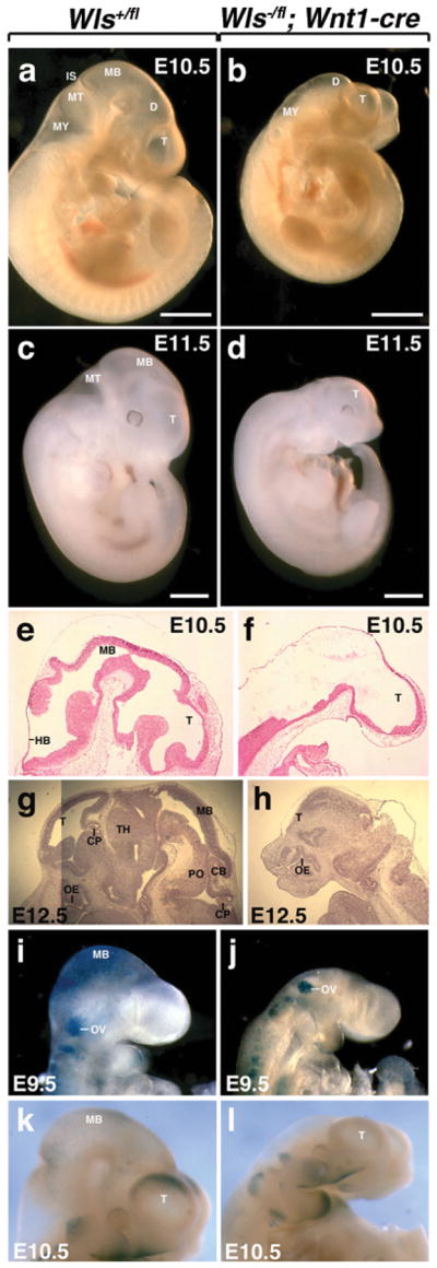
Deletion of Wls in the dorsal midline of the diencephalon using Wnt1-cre. (a, b) Morphological analysis of embryos dissected at E10.5. Wls+/fl embryos exhibit a forebrain, midbrain and hindbrain. Wls−/fl; Wnt1-cre embryos fail to develop a midbrain. (c) Wls+/fl embryos dissected at E11.5. (d) Wls−/fl; Wnt1-cre embryos at E11.5 show defective development of midbrain, hindbrain and forebrain. Histologic comparison of WT (e, g) and mutant embryos (f, h). Mutant embryos display loss of midbrain and hindbrain formation at E10.5 (f). At E11.5, mutant embryos additionally lack a forebrain choroid plexus and display a modified forebrain (h). β-catenin/Wnt activity in the brain of Wls+/fl (i and k) and Wls−/fl; Wnt1-cre (j and l) embryos stained for X-gal. Scale bars represent 1 mm. T, telencephalon; D, diencephalon; MB, midbrain; MT, metencephalon; MY, myencephalon; IS, isthmus; HB, hindbrain; CP, choroid plexus; OE, olfactory epithelium; TH, thalamus; PO, pons; CB, cerebellum; OV, otic vessicle.