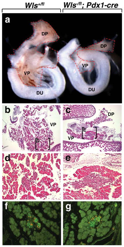
FIG. 4.
Deletion of Wls in pancreatic precursor cells using Pdx1-cre. (a, b) Morphological analysis of pancreata dissected at E15.5. Both Wls+/fl and Wls−/fl; Pdx1-cre pancreata exhibit a dorsal and ventral pancreas. Wls−/fl; Pdx1 pancreata are severely hypoplastic (b). Histologic comparison of WT (b, d, f) and mutant pancreata (c, e, g). Low-power magnification of both Wls+/fl and Wls−/fl; Pdx1-cre pancreata reveal the presence of acinar cells and Islets of Langerhans (b and c). Higher power magnification of the boxed regions in b and c (d and e, respectively). (f, g) E15.5 islets immunostained for insulin (green) and glucagon (orange). DP, dorsal pancreas; VP, ventral pancreas; Du, duodenum.

