Figure 3.

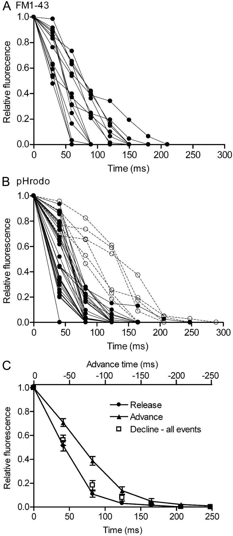
Fluorescence of dye-loaded organelles declined rapidly during depolarizing stimulation, consistent with fusion of individual vesicles. A, Overlay of fluorescence declines from 15 FM1–43-loaded vesicles. Vesicle fusion for FM1–43-loaded vesicles was defined as an abrupt decline in peak fluorescence intensity exceeding 50% within three frames (94 ms) with a total decrease of >90% relative to baseline fluorescence (filled circles, B). B, Overlay of fluorescence declines from 36 pHrodo-loaded vesicles. Vesicle fusion for pHrodo-loaded vesicles was defined as an abrupt decline in peak fluorescence intensity exceeding 60% within two frames (83 ms) with a total decrease of >90% relative to baseline fluorescence (filled circles, B). A few pHrodo-loaded vesicles (14%; 5 of 36) exhibited more delayed fluorescence declines (open circles, B) and were therefore not defined as fusion events. C, The average rate of fluorescence decline for single vesicles loaded with pHrodo (filled circles) was faster than the rise in fluorescence observed as vesicles approached the membrane (filled triangles), advancing deeper into the evanescent field of illumination. This difference was observed even when the few pHrodo-loaded vesicles that exhibited more delayed fluorescence declines (open circles, B) were included (open squares, C). When plotting the rate of fluorescence increase, the timescale was reversed (top ordinate) to facilitate comparison with rates of fluorescence decline.
