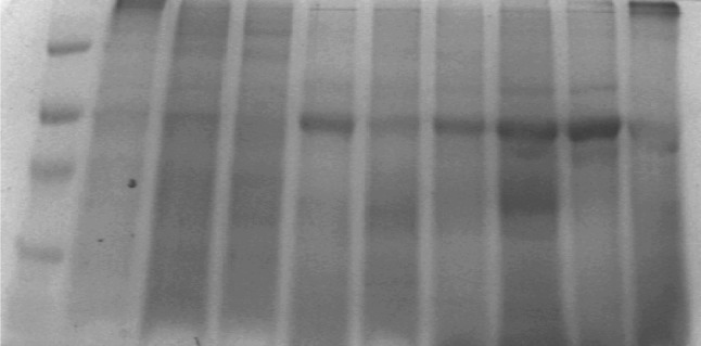
Fig. 3.

1. Marker; 2. Frontal (white); 3. Frontal (gray); 4. Cerebellum 40/M; 5. Pineal gland 40/M; 6. Pineal Gland 51/M, 7. Pineal gland 15/M; 8. Meningothelial meningioma G-I olfactory groove; 9. Anaplastic meningioma G-III recurrent right frontal 75/M; and 10. Glioblastoma multiforme (G-IV) frontal tumor shows florid micro vascular proliferation 40/M
