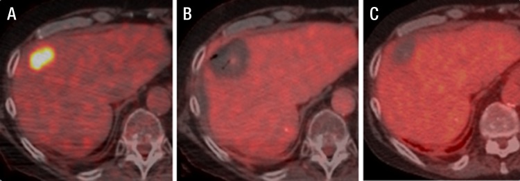
Figure 3:

Fused FDG PET/CT images in a 68-year-old man with metastatic colorectal cancer. A, Colorectal liver metastasis (standardized uptake value, 9.8). B, Intraprocedural image with no metabolic activity and decreased attenuation. C, Image of the same ablated area 4 months after the ablation.
