
Figure 5:
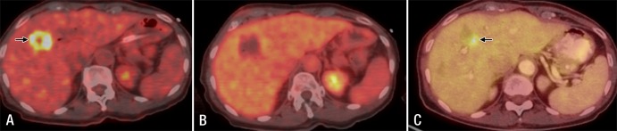
Fused FDG PET/CT images in a 69-year-old woman with metastatic pancreatic cancer. A, Image prior to ablation shows liver metastasis (arrow), which was treated with irreversible electroporation (not shown). B, Image at the end of ablation shows the ablation zone with no metabolic activity. C, Follow-up image at 3 months shows local tumor progression (arrow).