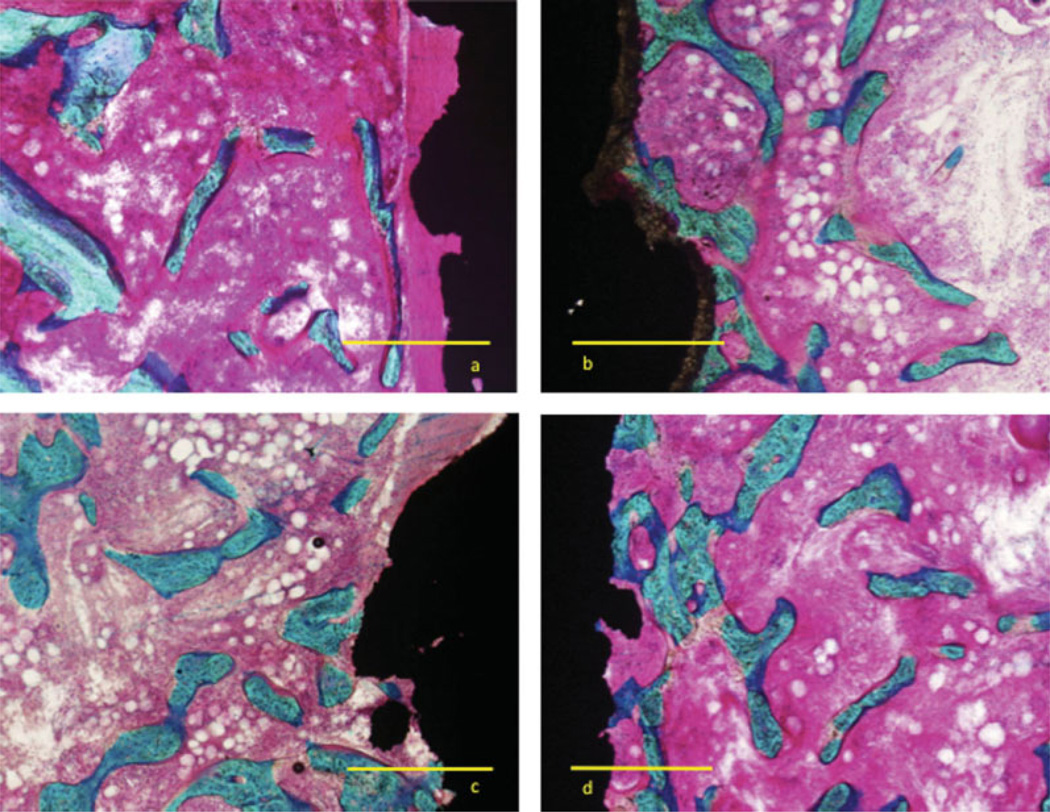
Figure 3.
Micrograph four weeks after insertion of plasma-sprayed titanium implant inserted in cancellous bone in 1-mm gap. Coating (a) plasma-spray titanium, (b) plasma-spray HA, (c) electrochemically deposited HA, and (d) electrochemically deposited HA mineralized collagen. Images showing area with bone ongrowth. Bar equals 500 µm. Basic fuchsine/Light Green stain. [Color figure can be viewed in the online issue, which is available at www.interscience.wiley.com.]

