Fig. 2.
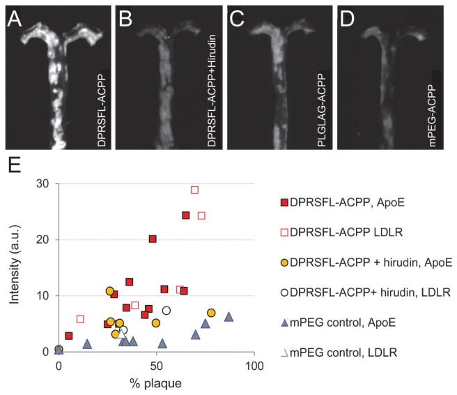
Fluorescent labeling of atherosclerotic aortas with thrombin or MMP cleavable ACPPs was enzyme dependent and correlated with increased plaque burden. (A–D) Representative fluorescence images are shown of gross aortas that were removed from ApoE−/− animals six hours after injection with either (A) 10 nmol DPRSFL–ACPP (n = 17 + 1 wildtype mouse); (B) 10 nmol DPRSFL–ACPP + hirudin (n=8+1 wildtype mouse); (C) 10 nmol PLGLAG–ACPP (n=11+1 wildtype mouse); (D) 10 nmol control mPEG–ACPP (n=9+1 wildtype mouse). Images A–D were acquired and processed using identical parameters. (E) Scatter plot showing average uptake of DPRSFL–ACPP in ApoE (closed shapes) and LDLR (open shapes) deficient mice as a function of plaque burden (quantification of data as shown in Fig. S1C, ESI†). Correlation coefficients for DPRSFL–ACPP, DPRSFL–ACPP + hirudin, PLGLAG–ACPP and mPEG–ACPP were 0.82, 0.48, 0.90 and 0.74 respectively. Slopes and 95% confidence intervals (in brackets) were 0.31 [0.19, 0.43], 0.06 [−0.04, 0.17], 0.15 [0.10, 0.21], 0.05 [0.03, 0.08] respectively.

