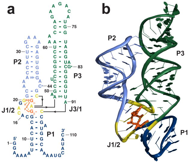
Figure 1.
c-di-GMP riboswitch from V. cholerae. a. Secondary structure of the riboswitch upstream of the tfoX-like gene in V. cholerae. The P1 helix is shown in dark blue, P2 in light blue, P3 in green. The asterisks next to C44 and G83 indicate that these residues are base paired. Nucleotides that directly contact the bases of c-di-GMP are show in yellow. c-di-GMP is shown in orange. b. Crystal structure. The U1A protein used for cocrystallization has been removed for clarity. Coloring is the same as in part a.

